How many members do you have on your team and have you ever
considered UniProt as one of them?
UniProt is a suite of open access protein databases, accessed by 9 million unique visitors a year, but how much money does it save you?
Our contribution to the scientific community and wider economy has now been analysed in a case study by CSIL, as part of the EU-funded project PathOS. This study investigated the cost-benefit of open data resources, and an analysis of UniProt’s impact between the years of 2017 and 2023 has been published.
What are the main costs of maintaining UniProt?
Across our three consortia sites, EMBL-EBI, SIB, PIR, we have office space, equipment, consumables and publication costs. But our main outgoings (70%) are salaries for our teams of expert biocurators and software developers. Cost to a user is measured in time spent providing valuable voluntary knowledge contributions. 49% of our users visit weekly and 26% daily.
What are the benefits to using UniProt?
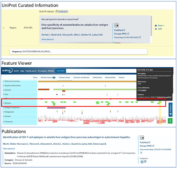
The immediate benefits for users are significant time savings. UniProt integrates data from over 180 cross-reference databases, combines it with expert manually curated sequence and protein data, then puts it all in one place, in a standardized format. This saves users time because they don’t have to navigate between multiple resources or download data in different formats. Additionally users do not have to sift through numerous scientific publications to understand the current cutting edge research available for a given protein. It also helps streamline research and analysis pipelines by providing centralized ready-to-use data.
Long-term benefits are measured in publications and patents that mention UniProt, indicating the wider impact on scientific knowledge advancement and technological development. Over a period of 7 years over 15,200 publications and over 183,000 patents cited or referenced UniProt. Many of these patents go on to be referenced by subsequent patents spanning a number of biotechnology and health innovation fields. Economic benefits have also been seen in the number of start-up companies that rely on UniProt data for their business model.

Demonstrating
the value of UniProt
- Each user gains a net benefit from
UniProt of up to €5,475 and saves 219 hours a year.
- Users say that our main strength ‘is the ability to integrate
protein sequences identified in the literature with an extensive
body of functional information’. This is facilitated by
incredibly passionate teams of expert biocurators and software developers
that ensure data is curated into the database reliably, and presented in a
consistent and easily accessible manner.
- Users agree ‘that there is no alternative offering the same breadth
of knowledge, quality and level of integration as UniProt’.
- Overall users say: ‘UniProt plays a crucial role in accelerating
scientific research and innovation in various forms, thereby facilitating
the creation of new knowledge.’
- In total UniProt provides a benefit of between €373-565 million per
year to its community of scientific users.
Learn more about the details of how UniProt supports research and innovation by reading the full report: Measuring the value and impact of open science
Links to associated articles
ELIXIR - https://elixir-europe.org/news/UniProt-CBA
SIB - https://www.sib.swiss/news/uniprot-user-benefits-up-to-39-times-higher-than-operational-costs
The UniProt team would like to take the opportunity to thank our funders, collaborators and users for their time, support and contributions to the database. UniProt is a key part of the EMBL-EBI, SIB Swiss Institute of Bioinformatics and the Protein Information Resource (PIR) and ELIXIR
infrastructure. Our main funders are EMBL, The State Secretariat for Education, Research and Innovation SERI (Switzerland), and NIH (USA). We are one of ELIXIR’s Core Data Resources, and two of our three partners, EMBL-EBI and SIB, are ELIXIR Nodes. The findings of this study will support efforts to advocate for long-term funding for critical biodata resources.